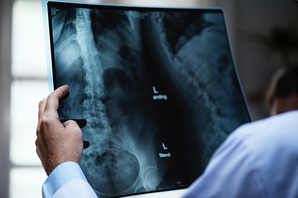
I never really considered the history and impact of X-rays, until my awareness grew around radiation exposure and its effects on children. I was fascinated and decided to do a bit of research on the subject, not knowing where this would lead.
Tim Newman posted an interesting article titled ‘Are X-rays really safe?’ on Medical News Today, 9 January 2018. Tim’s article was definitely one of the best I came across, it also helped me put a lot of the positive and the possible negative into perspective.
Did you know:
• A German mechanical engineer and physicist, Wilhelm Röntgen, was credited as producing and detecting electromagnetic radiation in a wavelength range known as X-rays or Röntgen rays, an achievement that earned him the first Nobel Prize in Physics in 1901.
• Just weeks after he discovered that they could help visualize bones, X-rays were being used in a medical setting.
• The first person to receive an X-ray for medical purposes was young Eddie McCarthy of Hanover, who fell while skating on the Connecticut
• River in 1896 and fractured his left wrist (https://www.ajronline.org/doi/pdf/10.2214/ajr.164.1.7998549).
• X-rays are a naturally occurring type of radiation.
• They are classed as a carcinogen.
• The benefits of X-rays far outweigh any potential negative outcomes.
• CT Scans give the largest dose of X-rays compared to other X-ray procedures.
• In X-rays, bones show up white, and gasses appear black.
• Everyone on the planet is exposed to a certain amount of radiation as they go about their daily lives. Radioactive material is found naturally in the air, soil, water, rocks, and vegetation. The greatest source of natural radiation for most people is radon.
• Additionally, the Earth is constantly bombarded by cosmic radiation, which includes X-rays. These rays are not harmless but they are unavoidable, and the radiation is at such low levels that its effects are virtually unnoticed.
• Pilots, cabin crew, and astronauts are at more risk of higher doses because of the increased exposure to cosmic rays at altitude.
• There have, however, been few studies linking an airborne occupation to increased incidence of cancer (I was not yet able to know if these studies are actually being conducted though…)
Importantly, and something that has been crossing my mind: Have you ever wondered how many x-rays and scans are safe in a lifetime? We all agree that in the world of medicine, science and technology, the most commendable accomplishments are medical imaging. The process of passing rays through the body to get images of the inside of the body, has helped doctors diagnose the severity of diseases with the right accuracy. X-rays, magnetic resonance imaging (MRI) and computerised tomography (CT) scans are the most prominent imaging technologies that we have at our disposal. However, the radiation that these imaging technologies pass through our bodies can impact our health.
All individuals are actually exposed to some sort of radiation every day. Natural background radiation comes from different things including the ground, air, food, and even from outer space in the form of cosmic radiation. Apart from this natural radiation we come into contact with every day, each x-ray we receive as well as nuclear medicine tests adds an additional dose to one’s exposure to radiation. The dose level of radiation varies depending on the medical examination done. X-ray exposure of the teeth, chest, and limbs usually have small radiation doses while exams involving more extensive use of x-rays like CT scans and fluoroscopy have higher radiation doses.
In my research, I found another interesting article by DoctorNDTV that helped me delve deeper into my questions: ‘Know all about the number of X-rays, MRI and CT scans you can get done in a lifetime to avoid risks of developing cancer’ – Updated: Mar 30, 2018
Studies indicate that maximum radiation risks are posed by CT scans – CT scans are done by an X-ray technique which is aided by the computer. Unlike the images produced by normal X-ray technique, CT scans give cross-sectional images of body parts and organs. Body’s exposure to radiation in CT scans is quite harmful.
X-rays are passed through the body. In order to get good quality image of the tissues, a dye or a contrast medium – made up of iodine or barium – is injected in the body. Since X-rays are passed through the body, they may pose a risk to our health because of exposure to radiation.
There is no radiation risk posed by MRIs. These scans work by using a magnetic field and radio waves which help in producing images of the internal structure of the body. The scan is done by creating a temporary magnetic field on a person’s body. The magnetic field is created by passing an electric current through coiled wires around the body. The transmitter or receiver sends and receives radio waves. The signals are used to produce scanned images of the body. Since MRI scan involves no radiation, it is a safe and pain-free process to scan any part of the body.
In the article, they ask Dr. Gita Prakash about how harmful these scans can be. She says:
“These scans do not increase the risk of cancer. You get them done because you worry about the risk of cancer. You have to be sensible about them and get them done only when needed. During pregnancy, you have to be careful that you don’t get any X-rays done and don’t expose the mother to any radiations.”
Measuring radiation in terms of natural radiation:
Low levels of ionising radiation are used to produce images in CT scans and X-rays. Ionising radiation is considered to be more harmful for the body as compared to non-ionising radiations.
The units in which we can measure radiation is known as millisieverts (mSv). Apart from the radiation through these imaging scans, our body is also exposed to radiation in the environment. The body is exposed to around 3.1 mSv of radiation through natural resources – states the United States Nuclear Regulatory Committee.
A single chest X-ray makes our body exposed to 0.01.4 mSv radiation. This is equivalent to 3 days of radiation from natural resources. An X-ray of the abdomen exposes our body to 0.7 mSv radiation, which is equivalent to 4 months of radiation through natural resources.
A CT scan of the head exposes our body to 2 mSv radiation, which is equivalent to 1 year of exposure through natural resources.
The next bit of the article was really where I realized it was not that simple at all – it asks the question: Can more CT scans increase your risks of cancer?
The safety of scans is determined by examining the dose of radiation as compared to the frequency of the scan. MRIs, as mentioned above, do not pose any risks of radiation.
Radiation exposure in a CT scan depends on the number of scans done, the patient’s size, the design of the scanner used, the time or rotation and/or exposure.
Risks of cancer depends on the age and sex of the patient, along with the type of scan and even type of scanner.
A radiation study says that of the 270 women who underwent CT coronary angiography at the age of 40, 1 can develop cancer from that CT scan. The CT scan will pose cancer risks to 1 in 600 men. A routine CT scan poses cancer risk to 1 in 8,100 women at the age of 40. A routine CT scan poses cancer risk to 1 in 11,080 men at the same age. The risks of developing cancer are double in people aged 20 years. For patients aged 60 years, the chances of cancer risks were 50% lesser. (Study: Radiation Dose Associated with Common Computed Tomography Examinations and the Associated Lifetime Attributable Risk of Cancer. Published in final edited form as: Arch Intern Med. 2009 Dec 14; 169(22): 2078–2086.)
The safety angle, according to experts, is that the damage done to the body, because of radiation done during a CT scan, is likely to get repaired within 1 year. This is because the radiation dosage is usually below the safe numbers. Nonetheless, it is still important for a person to understand the effects of radiation and take necessary steps to minimise exposure. (Dr Gita Prakash is a Family Physician at Max Multi Speciality Hospital, Panchsheel Park)
X-rays are believed to promote formation of free radicals in the body causing cell injury or cell death. Cells can either repair themselves with no damage done or there is also the possibility of cells improperly repairing themselves leading to changes in the cell’s structure and function. Reproductive organs, blood-forming organs, and digestive organs are considered to be the most sensitive to radiation while the muscle tissues, connective tissues, and the nervous system are through to be the least sensitive. The effects of radiation on the body depends on several factors including radiation dose, radiation energy, part of the body exposed, and cell sensitivity.
Common medical procedures that involve the use of x-rays usually have negligible to low risk. To be able to make an estimate of the likely effect of these examinations, you simply add the risks for each test together. It doesn’t make much of a difference if you have 10 chest x-rays this year, or if you have two chest x-rays per year over five years as the amount of x-ray radiation is what matters, and not the frequency.
It is clear in all my reading that awareness and knowledge is the greatest means to reducing exposure. We need to inform our doctors about X-rays or isotope scans we had in the past, so that unnecessary x-ray exposure from medical exams can be avoided. Parents need to be aware that risks of x-ray radiation exposure is greater in children, especially unborn babies.
X-rays can be valuable and have generally low health risks. Keeping track of medical examinations using x-rays that you have already undergone can be useful information for your doctor when deciding if more examinations should be avoided or not. In most cases, there is a higher risk from not having an x-ray examination or isotope scan compared to the risk of radiation itself. (Copyright: 2015, Anand Diagnostic Laboratory)
In a dedicated study around the effects of radiation in children, researchers followed 337 children under the age of 6, who had surgery for heart disease at Duke University Medical Center in North Carolina. The team, led by Dr. Kevin Hill, cardiologist and assistant professor of pediatrics at Duke, says they studied children with heart disease because they are exposed to more imaging tests than children in most other groups. The imaging procedures the children underwent totaled nearly 14,000. This includes X-rays, computed tomography (CT) scans and cardiac catheterization procedures using video X-rays – known as fluoroscopies.
Overall, the team found that the cumulative dose of ionizing radiation for the average child in the study was lower than the annual background exposure in the US. Though this finding can certainly put many parents’ minds at ease, the team did find that some children with complex heart disease, who are exposed to large cumulative doses of radiation have increased lifetime risks of cancer – up to 6.5% above baseline.
Commenting on their findings, Dr. Hill says:”There are definitely times when radiation is necessary. But it’s important for parents to ask and compare in case you can avert potentially high exposure procedures. Often there are alternative or modified procedures with less radiation, or imaging may not actually be necessary.” “Simple awareness is one of the greatest means to reducing exposure,” he says. “Health care providers should consider tweaking protocols to limit radiation doses and balance risks and benefits of every imaging study they do.” By Marie Ellis , Published Tuesday 10 June 2014
In 2013, Medical News Today reported on a study that suggested an anti-cancer compound present in cruciferous vegetables, such as cabbage, cauliflower and broccoli, protects rodents from radiation damage.
So, maybe that answer is more than we need, but we need to be more aware of what an X-ray is. Why it is needed? How does it compare to other tests for my condition? How much radiation is required? Answer these questions and weigh it against the benefits.
Far too long have we just accepted everything as fact – I never questioned a single scan, not for myself or my children.I never thought to ask questions about it, nor evaluated risk benefit . To be frank, the first time I started to really think about it, was when a friend’s physician opted to talk to her about not sending her young son for an x-ray, and took the time to explain to her the risk/benefit factors for young children. It was interesting to the both of us and hats off to that wonderful physician to take the time to bring this to our attention. It was only when I started to trawl through blogs, websites and studies that I realized the complexities.
Some practical take-aways:
The question: ‘How much medical radiation is too much?’ has no definitive answer.
A better question is: ‘How much radiation exposure is required to take care of my condition?’ Which will depend on your medical condition.
Ask your healthcare provider and radiologist about the benefits of the exam and the amount of radiation exposure involved. Ask your physician two questions: 1. What do we expect to learn from the x-ray exam? 2. Will decisions about my healthcare be determined from my imaging exam? You and your healthcare provider must work together to decide what is necessary and best for you.
One follow-up question to ask might be: Is an x-ray, CT scan or nuclear imaging exam the best exam, or would other exams like ultrasound, magnetic resonance imaging (MRI) or lab tests, work as well? Your physician can then explain the different examinations, their benefits, risks and results in line with your condition.
Asking questions can help us understand why we need an imaging exam and which one is best for our healthcare. Effective access to our health records also allow less duplication of tests and the sharing of critical information and patient education so that we can take better control of our health and healthcare in general.
Writer – Business Development Team


